Microscopic Imaging Core

Wake Forest Biology Department

Menu
  • Home
  • Instrumentation
    • AxioZoom V16
    • Inverted Microscope
    • Leica Thunder Live
    • LSM 880 Confocal Microscope
    • Microscopes across campus
    • Nikon SMZ25 Stereoscope
    • Revolve4 Microscope
    • Scanning Electron Microscope
    • Upright Microscope
    • LSM 710 Confocal Microscope
  • News and Events
    • Publications
  • Acquisition
    • Acquisition and Analysis Protocols
  • Writing Help
    • Grant Help
    • Publications and Acknowledgments
  • Resources
    • Book Resources
    • Electronic Resources
    • Ethical Image Editing
    • Free Analysis Software
    • Sample Preparation
  • Contact

Author: Facility Director

Covid-19 Operational Guidelines

July 12, 2020
| No Comments
| Updates

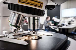
Wake Forest Microscopy Core Covid-19 Operational Guidelines Adoption of these new practices and guidelines are intended to limit physical interactions and promote a safe work environment for all microscopy facility users.  Updated Regulations and Distancing Rules: 24 hour advanced electronic equipment signup/scheduled strongly encouraged (no drop-in usage) Imaging sessions should include 30 minute gaps  to…
Read More »

Posted in Updates

Conner Lab Uses SEM for Publication

January 15, 2020
| No Comments
| publications

The Conner Lab recently published an article titled “Characteristics of tiger moth (Erebidae: Arctiinae) anti-bat sounds can be predicted from tymbal morphology.”  In this article, the facilities Amray 1810 SEM microscope was used to image the thorax of tiger moths.  Undergraduate researchers Erika Metzler, James Clemmons, Thisbe Scholfield-Johnson, and Kahla Seymour participated in obtaining the SEM…

Read More »

Posted in publications

Lukesh Lab develops dyes for lipids and H2S

January 15, 2020
| No Comments
| publications

At  Wake Forest we pride we pride ourselves on the wonderful collaborative environment between departments.  In the imaging field we move forward with collaboration between biologists, chemists, physicists, engineers, and even computer scientists.  The Lukesh Lab has recently published two papers on fluorescent probes they have developed that allow biologists to visualize lipid droplets and…

Read More »

Posted in publications

Microscopy Teaching Lab

August 1, 2019
| No Comments
| Updates

  This spring, we opened the doors to the new microscopy teaching lab!  The microscopy teaching center (MTC) has been a long term goal of the core with Dr. Glen Marrs beginning the process approximately five years ago.  Typically, teaching labs are set up with old clunky microscopes, which are challenging to use and only…

Read More »

Posted in Updates

Publications for FY2019

July 31, 2019
| No Comments
| publications

This year the core has been quite busy, and many papers have been published!  Check out all the work that has been done! J. K. Muhlemann, T. L. B. Younts, G. K. Muday, Flavonols control pollen tube growth and integrity by regulating ROS homeostasis during high-temperature stress. PNAS. 115, E11188–E11197 (2018). E.R. Shuppe, M.J. Fuxjager,…

Read More »

Posted in publications

Johnson Lab GPCR paper

August 22, 2018
| No Comments
| publications

The Johnson Lab recently published “Unexpected role of a conserved domain in the first extracellular loop in G protein-coupled receptor trafficking.”  The lead author was graduate student Michael Rizzo who performed his experiments on the Zeiss 710 confocal microscope in the Winston Hall microscopy core.  Also involved in this study were undergraduate researchers John “Jack”…

Read More »

Posted in publications

FY 2018 Publications

July 11, 2018
| No Comments
| publications

We keep statistics for all sorts of things from July 1- June 30th every year.  One of the most important statistic for us is the number and type of publications that come from researchers utilizing our core facility.  We are hoping with the second site Downtown, 3 more pieces of equipment, and new hires we…

Read More »

Posted in publications

New Fluorescent Microscope

May 2, 2018
| No Comments
| new events

  This week we had new fluorescent microscope installed.  It is called the Echo Revolve microscope. (https://discover-echo.com) It can be used as an inverted microscope (samples in dishes) or as a upright microscope (slides, material samples).  Also, the Muday lab will be using it in a horizontal position to perform gravitropic experiments. The microscope has:…

Read More »

Posted in new events

Image Analysis Workshop

February 22, 2018
| No Comments
| event

  Looking to take your data up to the next level?  Join us for an image analysis workshop put on by Aivia on 2/28 9-10 Introduction to Image Analysis 10-12 Try out Virtual Reality 1-5 Bring your images and have Aivia help you with analysis Click here to register!

Read More »

Posted in event

Airyscan “how to” up in Resources

November 8, 2017
| No Comments
| Resource

We have recently created some “cheat sheets” to help our users on the confocal microscope. They, along with other techniques can be found in Acquisition & Analysis. Airyscan Handbook  & Airyscan Processing

Read More »

Posted in Resource
  • 1 of 3
  • 1
  • 2
  • 3
  • Next »

Follow us!

Follow Us on TwitterFollow Us on Facebook

Recent Posts

  • Covid-19 Operational Guidelines
  • Conner Lab Uses SEM for Publication
  • Lukesh Lab develops dyes for lipids and H2S
  • Microscopy Teaching Lab
  • Publications for FY2019

Recent Posts

  • Covid-19 Operational Guidelines
  • Conner Lab Uses SEM for Publication
  • Lukesh Lab develops dyes for lipids and H2S
  • Microscopy Teaching Lab
  • Publications for FY2019

Recent Posts

  • Covid-19 Operational Guidelines
  • Conner Lab Uses SEM for Publication
  • Lukesh Lab develops dyes for lipids and H2S
  • Microscopy Teaching Lab
  • Publications for FY2019

Recent Posts

  • Covid-19 Operational Guidelines
  • Conner Lab Uses SEM for Publication
  • Lukesh Lab develops dyes for lipids and H2S
  • Microscopy Teaching Lab
  • Publications for FY2019

Wake Forest University
Microscopy Imaging Core
Winston Hall

  • Home
  • Instrumentation
    • AxioZoom V16
    • Inverted Microscope
    • Leica Thunder Live
    • LSM 880 Confocal Microscope
    • Microscopes across campus
    • Nikon SMZ25 Stereoscope
    • Revolve4 Microscope
    • Scanning Electron Microscope
    • Upright Microscope
    • LSM 710 Confocal Microscope
  • News and Events
    • Publications
  • Acquisition
    • Acquisition and Analysis Protocols
  • Writing Help
    • Grant Help
    • Publications and Acknowledgments
  • Resources
    • Book Resources
    • Electronic Resources
    • Ethical Image Editing
    • Free Analysis Software
    • Sample Preparation
  • Contact

© Wake Forest University